Cranioplasty: how the integrity of the skull is restored after serious injuries
Cranioplasty is the surgical repair of defects in the cranial vault
(the dome-shaped part of the skull). It sounds complicated, but in
reality, it is an operation that closes the “hole” in the head
caused by trauma or disease. Why is this necessary? First, the brain
needs to be protected from mechanical damage. Second, an intact skull
helps fluids circulate properly around the brain. Third, it is
important for the patient's appearance and psychological comfort.
When is cranioplasty
necessary?
Skull defects can
occur in several ways:
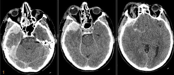
After severe head
injuries. In cases of serious fractures, when the bone is shattered,
surgeons have to remove the damaged fragments to prevent them from
entering the brain.
In cases of critical
brain edema. When the brain swells sharply due to a stroke,
hemorrhage, or severe trauma, doctors perform a decompressive
craniectomy — removing part of the bone to give the brain room to
expand and save the patient's life. Later, when the swelling
subsides, this bone must be put back in place.
Due to infection.
Sometimes an infection of the bone tissue (osteomyelitis) develops in
the area of the operation, and the infected area of the bone has to
be removed.
For tumors. If a
tumor affects the skull bone, surgeons remove that area along with
the tumor and then restore the brain's protection.
Trepanation
syndrome: when the defect affects life
After removing part
of the skull, patients often develop an unpleasant condition that
doctors call trepanation syndrome. Imagine: a hollow forms in the
head, and the skin above it sinks inward. Atmospheric pressure
literally presses on the brain through this hollow.
How it feels:
-- Headache —
often worse when standing or sitting. The pain is less severe when
lying down because atmospheric pressure is less intense.
-- Dizziness and
fatigue — patients feel exhausted and have difficulty
concentrating.
-- Memory and
thinking problems — the person has trouble remembering information,
finds it difficult to plan, and becomes distracted.