

creator cover Neurosurgeon, Ph.D. Sergey Tikhomirov
Neurosurgeon, Ph.D. Sergey Tikhomirov

Neurosurgeon, Ph.D. Sergey Tikhomirov 

Neurology & Neurosurgery. Online consultation.

0subscribers

83posts

About

pdf
Направление МРТ-МСКТ физ_лица.pdf331.87 Kb

Chronic Subdural Hematoma: Why It Occurs and How It Is Treated

A minor bump to the head does not always seem like a serious event. But sometimes, several weeks later, a person develops a headache, unsteadiness, or unusual drowsiness. The cause may turn out to be a chronic subdural hematoma — a collection of blood and fluid beneath the dura mater.
This condition most often affects older adults. Its hallmark is that the hematoma can grow slowly, and the link between symptoms and a long-past injury is not always obvious. At the same time, modern research shows that the problem is not only about a one-time bleed. A prolonged process of inflammation and formation of fragile vessels can be sustained around the hematoma.

What Happens Inside the Skull

The brain is surrounded by several membranes. One of them is the dura mater. When tissues near it are damaged, a cavity can form between the membranes, where blood and other fluid accumulate. If the collection persists and develops gradually, it is called a chronic subdural hematoma, or CSDH.
The triggering event is often a fall or a blow to the head. However, the injury can be so minor that the person does not remember it. Sometimes no clear cause can be identified.
The risk of CSDH is increased by:
  • older age;
  • reduced brain volume, which makes the vessels between the brain and its membranes more vulnerable to stretching;
  • use of anticoagulants and antiplatelet agents — drugs that reduce the blood's ability to clot;
  • repeated falls and certain co-existing conditions.
The presence of risk factors does not mean that a hematoma will necessarily develop, but it helps the physician assess the likelihood of the disease.

Why the Hematoma Can Grow

CSDH used to be often described as a passive collection of old blood. Today, researchers view it as a more complex process.
After tissue damage, inflammation develops in the area of the hematoma. Thin membranes may form around the accumulated fluid. New blood vessels appear in the outer membrane, but their walls are often fragile and permeable. Because of this, fluid and small amounts of blood can re-enter the hematoma cavity.
At the same time, processes that break down formed blood clots intensify within it. As a result, fresh bleeding may be harder to stop.
This creates a kind of vicious circle: inflammation promotes the formation of fragile vessels, and repeated microbleeds sustain inflammation and hematoma growth. This is one reason why it sometimes enlarges long after the initial injury and can recur after treatment.
Chronic Tension-Type Headache: Why "Ordinary" Pain Can Become Daily and How to Overcome It
Post is available after purchase
Binswanger's Disease: The Silent Destruction of the Brain's "Wiring"
Post is available after purchase
Migraine: What Happens in the Brain and How to Cope with It
Post is available after purchase
Diffuse Axonal Injury: A Guide for Patients and Families from Diagnosis to Rehabilitation
Post is available after purchase

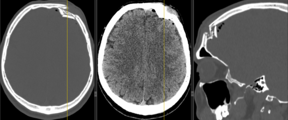
Plastic repair of the cranial vault defect in case of a depressed fracture.

Patient B., 39 years old, was taken by an ambulance from Rodnikovskaya Central District Hospital to Kineshemskaya Central District Hospital. The injury was caused by hitting his head on a metal sheet.
He underwent an X-ray examination and then a CT scan of the brain. A depressed penetrating fracture of the frontal bone was detected.
MSCT of the brain performed upon admission.

Cranioplasty: how the integrity of the skull is restored after serious injuries

Cranioplasty is the surgical repair of defects in the cranial vault
(the dome-shaped part of the skull). It sounds complicated, but in
reality, it is an operation that closes the “hole” in the head
caused by trauma or disease. Why is this necessary? First, the brain
needs to be protected from mechanical damage. Second, an intact skull
helps fluids circulate properly around the brain. Third, it is
important for the patient's appearance and psychological comfort.
When is cranioplasty
necessary?
Skull defects can
occur in several ways:
After severe head
injuries. In cases of serious fractures, when the bone is shattered,
surgeons have to remove the damaged fragments to prevent them from
entering the brain.
In cases of critical
brain edema. When the brain swells sharply due to a stroke,
hemorrhage, or severe trauma, doctors perform a decompressive
craniectomy — removing part of the bone to give the brain room to
expand and save the patient's life. Later, when the swelling
subsides, this bone must be put back in place.
Due to infection.
Sometimes an infection of the bone tissue (osteomyelitis) develops in
the area of the operation, and the infected area of the bone has to
be removed.
For tumors. If a
tumor affects the skull bone, surgeons remove that area along with
the tumor and then restore the brain's protection.
Trepanation
syndrome: when the defect affects life
After removing part
of the skull, patients often develop an unpleasant condition that
doctors call trepanation syndrome. Imagine: a hollow forms in the
head, and the skin above it sinks inward. Atmospheric pressure
literally presses on the brain through this hollow.
How it feels:
-- Headache —
often worse when standing or sitting. The pain is less severe when
lying down because atmospheric pressure is less intense.
-- Dizziness and
fatigue — patients feel exhausted and have difficulty
concentrating.
-- Memory and
thinking problems — the person has trouble remembering information,
finds it difficult to plan, and becomes distracted.

Kimmerli's anomaly

Kimmerli's anomaly, also known as ponticulus posticus, is an anatomical variant involving the ossification of the posterior atlanto-occipital membrane over the groove of the vertebral artery, leading to the formation of an arcuate foramen or ridge (). This anomaly can result in extravasal compression of the vertebral artery, which can sometimes cause cerebrovascular symptoms.
A complete Kimmerli anomaly on the right and an incomplete one on the left.

Head wound from an air gun.

Patient C., 16 years old, applied. According to the patient's words, the previous day he accidentally shot himself in the head from a pneumatic weapon. On visual examination, an entrance hole with a diameter of 2-3 mm was seen in the right frontal region.
Intraoperative photograph. The entrance hole in the frontal region on the right side is visible.
The patient underwent MSCT of the brain. A foreign body is seen in the soft tissues of the frontal region. Due to metal artifacts it is not possible to make an unambiguous conclusion about the presence or absence of damage to the frontal bone.
MSCT of the brain: axial and coronal scans show a metallic foreign body. Due to artifacts, it is difficult to clearly determine whether or not there is a frontal bone lesion.
Subscription levels0
No subscription levels
Go up